Standard return-to-play protocols operate under a flawed assumption: that if an athlete reports no lingering headaches or brain fog, they are cleared for competition. In elite athletics and tactical environments, this standard fails. Most head injuries disrupt the visual system in ways standard cognitive baselines miss completely. Medical staff routinely clear players when basic symptoms resolve, leaving underlying ocular-motor dysfunction intact.

These unseen vision deficits directly impair performance. When an athlete’s eyes cannot converge on a moving target, adjust focus across distances, or track rapid movements, the brain burns massive metabolic energy just trying to parse visual input. Layer on game-film study, complex tactical reads, or high-speed field decisions, and this deficit causes cognitive overload.
Unaddressed visual issues lead to persistent motion sensitivity, headaches, nausea, and anxiety. Because vision is rarely evaluated during standard rehab, these issues get mislabeled as generalized post-concussion syndrome or stress. The result is extended recovery timelines, degraded performance, and a higher risk of secondary injury upon returning to play.
To protect athletes and optimize return-to-play infrastructure, performance directors and medical staff must address visual-motor rehab with the same objective rigor used in musculoskeletal recovery.
The Physiology of Post-Concussive Visual Breakdown
The visual system is an active extension of the central nervous system. Over 50% of neural pathways involve visual processing, direct motor output, and spatial orientation (Master et al., 2016). When impact forces shear axons or trigger a neurometabolic cascade, they disrupt the visual-vestibular network that keeps visual fields stable during movement.
Clinical research shows that up to 82% of post-concussion patients present with measurable ocular-motor dysfunction (Ciuffreda et al., 2017; Master et al., 2016). Visual impairment is not an edge-case symptom; it is the dominant manifestation of central sensory mismatch.
The Three Primary Subsystems
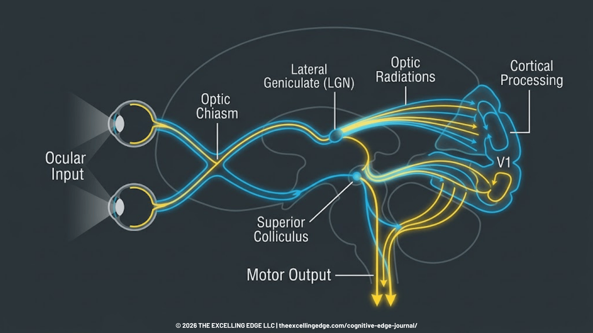
- Visual Convergence: The inward rotation of both eyes to maintain single focus on a near object, controlled by Cranial Nerve III. Post-concussion, convergence insufficiency occurs in up to 55% of cases. One eye drifts outward under fatigue, causing blurred or double vision and disrupting depth perception.
- Accommodation: The eye’s ability to adjust lens focus across distances. Accommodative breakdown, identified in up to 65% of concussive injuries, prevents an athlete from rapidly switching focus from a far target (surveying the field) to a near target (tracking a ball into their hands).
- Saccadic Eye Movements: Fast, precise eye movements that switch fixation between targets. Saccades rely on the frontal eye fields, superior colliculus, and brainstem (Mucha et al., 2014). Trauma causes saccadic dysmetria—overshooting or undershooting visual targets—which slows overall processing speed.
The Metabolic Cost of Visual Dysfunction
When these subsystems fail, the brain receives conflicting signals from the eyes and inner ear. To compensate, the brain reallocates processing power simply to keep the visual environment stable. This constant correction drains central nervous system reserves, explaining why concussed athletes experience sudden fatigue or headaches when reading, analyzing film, or navigating crowded venues (Kontos et al., 2017).
Lab Notes: The Hidden Metabolic Drain
Misaligned ocular-motor systems force the brain to treat visual blur (retinal slip) as a constant error. The continuous neurological effort to correct this error causes rapid central nervous system fatigue, triggering headaches and dizziness during basic visual tasks.
High-Stakes Application: UFC Return-to-Sport Case Analysis
At our Neuro Performance Center in Surprise, AZ, we evaluate athletes, tactical operators, and active aging clients who have dealt with lingering post-concussion symptoms for months or years after an injury.
We were called in to assist with the return-to-sport protocol for an active UFC fighter. His physical therapist worked to bring his resting symptoms back to baseline. However, whenever under physiological activation, he experienced subtle spatial disorientation and timing delays that left him open to counter-strikes. Traditional concussion batteries marked him as fully recovered, but his functional tracking told a different story.
Using digital diagnostics and sensory station technology, we evaluated his ocular systems under movement. We identified an isolated visual field deficit and a clear delay in neuro-motor reaction time. His saccadic latency—the time required to spot a strike and initiate a visual response—was significantly degraded.
Clearing him based on symptom reports alone would have placed him at extreme risk for a secondary head injury. We built a visual-motor integration protocol alongside his PT, combining strobe eyewear with real-time heart-rate tracking to match the conditioning demands of a fight. By restricting visual information while pushing cardiovascular load, we trained his visual cortex to process sparse data faster.
Within weeks, re-testing showed complete recovery of his saccadic speed and visual-motor integration. He returned to sparing and competing with full spatial awareness, quicker visual processing and reaction speed.
The Return-to-Play Framework: The Four-Phase Visual-Motor Protocol
Visual-motor deficits are both measurable and trainable. You can integrate this four-phase framework directly into existing rehabilitation pipelines.
Phase 1: Objective Baselines and VOMS Screening
Establish an objective baseline using the Vestibular/Ocular Motor Screening (VOMS) alongside standard cognitive baselines like ImPACT.
- Action: Test smooth pursuit, horizontal and vertical saccades, Near Point of Convergence (NPC), and the Vestibulo-Ocular Reflex (VOR).
- Metric: A normal NPC is under 5 cm. Any distance beyond 5 cm, or any symptom provocation (nausea, dizziness, eye strain) during saccades, signals functional visual disruption.
Phase 2: Ocular-Motor Isolation
Isolate and retrain the cranial nerve pathways responsible for convergence and tracking once resting symptoms are stable.
- Action (Brock String Drills): Use a 10-foot Brock String with multi-colored beads to train convergence and binocular alignment.
- Execution: Have the athlete focus on the nearest bead, confirming both eyes align on it (forming a clean visual ‘X’), then rapidly shift focus to middle and far beads. Hold focus for 3 seconds per target to rebuild ciliary muscle control.
Phase 3: Visual-Motor Integration & Cognitive Loading
Evaluating cognitive-motor function in isolation creates false positives. Studies show that athletes cleared by standard static baselines still experience degraded reaction times and visual-motor breakdowns under dual-task condition loading—elevating secondary injury and concussion risk by up to 300% upon return to play (Brooks et al., 2016).
- Action (Strobe Eyewear Protocol): Introduce stroboscopic glasses during footwork, catching drills, or tactical movements to simulate field conditions.
- Execution: Perform dynamic movement and sport-specific while the eyewear alternates between clear and opaque states. Removing milliseconds of visual feedback forces the nervous system to predict target trajectories and process visual input faster under cognitive load.
Phase 4: Autonomic Stress Integration
Re-evaluate visual processing under high heart rate conditions. Visual deficits frequently re-emerge when the sympathetic nervous system is engaged (Howell et al., 2013). Active, sub-symptom threshold aerobic exercise is proven to speed recovery by restoring autonomic nervous system control and normalizing cerebral blood flow.
- Action: Combine high-intensity cardiovascular efforts (e.g., Assault Bike sprints) with dynamic target tracking or call-out visual charts or light targets.
- Metric: Clear the athlete only when visual processing speed and accuracy match baseline levels under maximum cardiovascular exertion.
Lab Notes: Multi-Disciplinary Standard
Vision training does not replace physical therapy; it completes it. Effective return-to-play requires physical therapists, athletic trainers, and neuro-performance staff working from the same objective dataset.
Ensure Complete Competitive Readiness
Relying on subjective symptom checklists and basic pen-and-paper baselines leaves performance on the table and puts athletes at risk. If your return-to-play pipeline does not measure and train visual-motor processing—under physiological and cognitive load, your clearance protocol is incomplete.
Book a 15-Minute Neuro-Tech Audit Call to review your organization’s RTP process and ensure the long-term health and safety of your athletes and operators.
References
- Brooks, M. A., Peterson, K., Biese, K., Sanfilippo, J., Heiderscheit, B. C., & Bell, D. R. (2016). Concussion increases odds of sustaining a lower extremity musculoskeletal injury after return to play among collegiate athletes. The American Journal of Sports Medicine.
- Ciuffreda, K. J., Ludlam, D. P., & Yadav, N. K. (2017). Conceptual model of optometric vision therapy in mild traumatic brain injury (mTBI). Vision Development & Rehabilitation.
- Howell, D. R., Osternig, L. R., van Donkelaar, P., Mayr, U., & Chou, L. S. (2013). Effects of concussion on attention and executive function in adolescents. Medicine and Science in Sports and Exercise.
- Kontos, A. P., Deitrick, J. M., Collins, M. W., & Mucha, A. (2017). Review of vestibular and oculomotor screening and concussion rehabilitation. Journal of Athletic Training.
- Master, C. L., Scheiman, M., Gallaway, M., et al. (2016). Vision diagnoses are common after concussion in adolescents. Clinical Pediatrics.
- Mucha, A., Collins, M. W., Elbin, R. J., Furman, J. M., et al. (2014). A brief vestibular/ocular motor screening (VOMS) assessment to evaluate concussions: preliminary findings. The American Journal of Sports Medicine.
Please note: I encourage reader discussion, however, I reserve the right to delete comments that are offensive or off-topic.