Non-contact musculoskeletal (MSK) injuries—specifically anterior cruciate ligament (ACL) tears, medial collateral ligament (MCL) sprains, and high-ankle sprains—cost professional and collegiate sports organizations hundreds of millions of dollars annually in lost roster value, rehabilitation costs, and competitive draft capital (Swanik et al., 2007; Swanik et al., 2017).

For decades, sports medicine and high-performance programs have approached injury prevention through a mechanical lens: measuring eccentric hamstrings strength, optimizing jump-landing mechanics, monitoring acute-to-chronic workload ratios, and training single-leg stability. Despite these rigorous physical protocols, non-contact ACL injuries account for approximately 70% to 80% of all ACL ruptures in multidirectional sports such as soccer, basketball, and football.
This persistent failure points to a missing link in high-performance conditioning: non-contact joint collapse is rarely a primary strength failure. It is a sensory-motor timing error.
An ACL tear occurs within 30 to 50 milliseconds after foot strike—far faster than the reflex loops of the spinal cord or brainstem can execute reactive corrections. When an athlete decelerates, cuts, or lands under mid-season physical and cognitive fatigue, even a 50-millisecond delay in visual processing destabilizes lower-extremity biomechanics. The nervous system fails to anticipate ground reaction forces, motor units fire out of sequence, and the knee collapses into high-risk dynamic valgus before the brain registers foot-ground contact.
Preventing non-contact MSK injuries requires shifting from isolated biomechanical screening to dual-task neurocognitive loading. By forcing the central nervous system to maintain dynamic joint stability while executing rapid visual decision-making, performance staff can build high-load cognitive resilience into dynamic movement.
The Biological Mechanism: Sensory-Motor Integration Under Cognitive Load
To understand why a healthy knee collapses without physical contact, we must analyze the visual processing loop and feedforward motor control.
Visual-Spatial Processing and Feedforward Motor Control
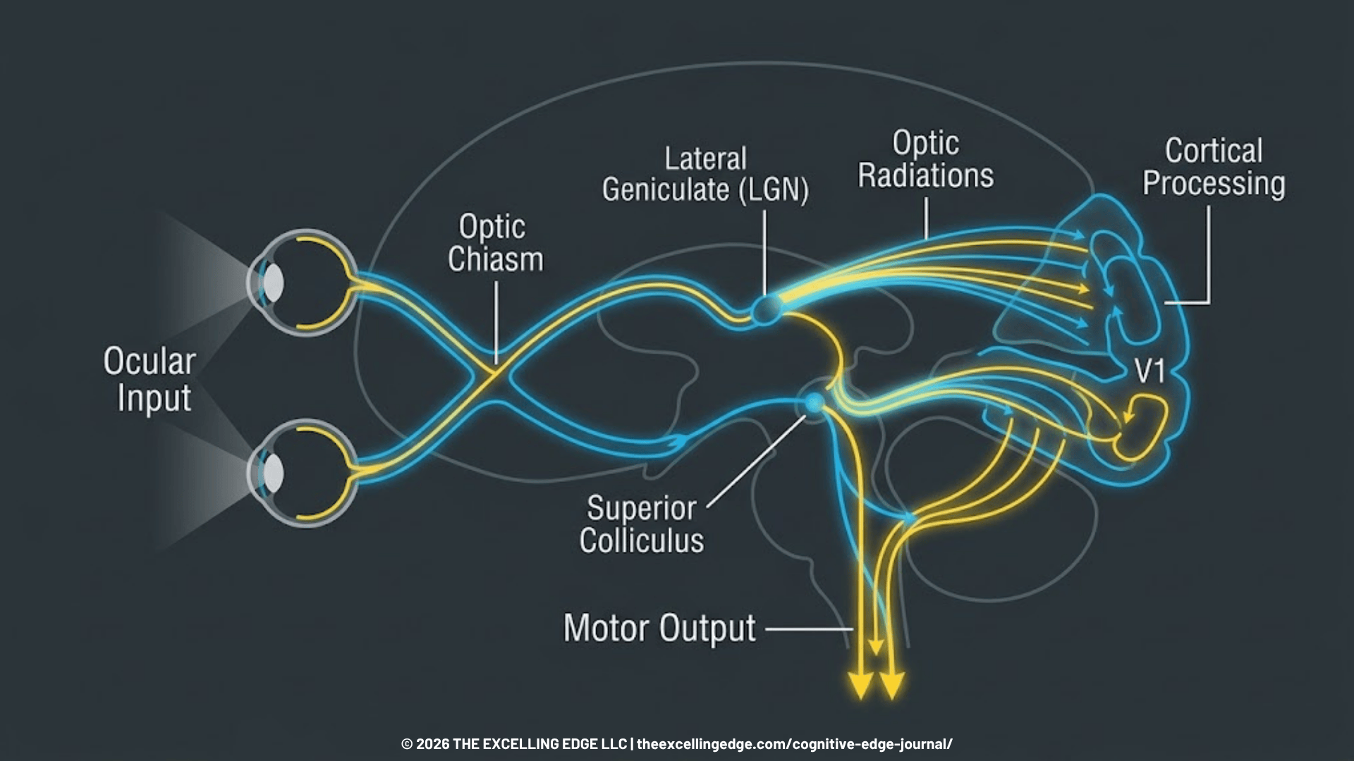
Athletes do not move in a vacuum; they navigate dynamic environments requiring continuous visual scanning. Visual information travels from the retina to the primary visual cortex before processing through two distinct pathways:
- Ventral Stream (“What” pathway): Decodes object identification and fine detail.
- Dorsal Stream (“Where/How” pathway): Computes spatial awareness, motion detection, and visual-motor transformations within the parietal cortex.
The dorsal stream feeds directly into the premotor cortex and supplementary motor area (SMA) to establish feedforward motor control. Because active muscle activation requires time to generate joint stiffness, the central nervous system (CNS) cannot rely purely on feedback mechanisms during rapid change-of-direction maneuvers. Pre-activation of the quadriceps, hamstrings, and gluteal musculature must occur prior to initial foot-ground contact.
The Cascade of Visual Processing Delays
When an athlete experiences an unexpected stimulus—such as a defender’s sudden step, a deflected pass, or an altered trajectory—the brain must execute rapid visual decision-making.
Research demonstrates that athletes who suffer non-contact ACL injuries exhibit baseline deficits in visual reaction time, visual processing speed, and cognitive flexibility compared to uninjured controls (and individual baseline data).

- Cognitive Latency: Under heavy cognitive load or physical fatigue, processing a visual stimulus slows down by 50 to 100 milliseconds.
- Attenuated Pre-Activation: The delayed visual processing signal fails to generate timely motor activation commands.
- Loss of Dynamic Restraint: The athlete strikes the ground with insufficient hamstring co-contraction, excessive knee extension, and elevated hip adduction.
- Mechanical Failure: Peak ground reaction forces (often exceeding 4–5 times body weight) load the ACL beyond its mechanical limit within 50 milliseconds of impact—long before spinal reflexes can intervene.
Lab Note: The 50-Millisecond Window
- Incidence Speed: Non-contact ACL rupture occurs within 30–50 ms post-contact.
- Feedback Latency: Spinal reflex loops take 80–120 ms to generate protective muscle torque; cortical motor commands take 150–250 ms.
- Mechanism: Protective joint stabilization relies entirely on feedforward motor planning driven by accurate visual input prior to initial ground contact.
Real-World Application: Bridging Academic Research and Elite Performance
At the Neuro Performance Center in Surprise, AZ, we bridge the gap between academic neuroscience and field execution. During a recent collaboration with an NFL franchise, the primary objective was clear: reduce non-contact lower-extremity injury risk while maximizing on-field agility. To achieve this without increasing the athletes’ physical workload, we anchored the team’s protocols in established neurocognitive research benchmarks.
The Research Benchmark: Why Cognitive Load Matters
Clinical data consistently demonstrates that physical strength alone cannot prevent joint failure if the nervous system cannot process the environment fast enough.
Swanik et al. (2007) established a foundational benchmark: athletes demonstrating lower baseline neurocognitive performance—specifically in visual processing speed and reaction time—experience significantly higher rates of non-contact ACL injuries. Furthermore, research by Grooms et al. (2016) highlights that the brain relies heavily on visual feedback to maintain motor control during dynamic movement.
When an athlete experiences mid-season metabolic fatigue, their visual processing speed inherently slows down. If the brain cannot process visual spatial data in real-time, feedforward motor planning is delayed, leaving the joint mechanically unsupported at the moment of foot strike.
Practical Implementation: The Living Lab Approach
To operationalize this research, we do not treat cognitive training as a separate, isolated exercise. Instead, we embed neurocognitive demands directly into standard biomechanical training flows.
During baseline evaluations with elite athletes, we consistently observe a distinct clinical pattern: under heavy physical exertion, visual reaction times degrade substantially. In high-speed change-of-direction tasks, this cognitive latency immediately manifests as compromised landing mechanics, delayed foot placement, and increased dynamic valgus stress.
To systematically close this gap, we integrate dual-task neurocognitive loading into the athletes’ existing routines:
- Visual Pre-Activation (Stroboscopic Integration): Prior to field sessions, athletes utilize stroboscopic eyewear during standard warm-ups (e.g., agility ladders, medicine ball tosses). By intermittently restricting visual input, the nervous system is forced to process spatial data more efficiently, priming the dorsal visual stream before high-velocity movement.
- Open-Loop Decision Making: We convert pre-planned cone drills into open-loop reactive tasks. Athletes must execute high-speed deceleration or directional cuts based strictly on randomized visual stimuli (e.g., light arrays or directional target boards) rather than auditory whistles or predetermined routes.
- Inhibitory Control Under Fatigue: Late in the conditioning session, athletes are tasked with executing plant-and-cut maneuvers only when specific visual criteria are met. This forces the central nervous system to maintain executive function and response inhibition even when heart rate and blood lactate levels are peaking.
Lab Note: Pre-Planned vs. Open-Loop Agility
- The Flaw of Pre-Planned Drills: Traditional agility drills allow the brain to pre-program the movement sequence. This tests force production but entirely bypasses visual decision-making.
- The Open-Loop Advantage: Forcing an athlete to react to unpredictable visual cues replicates game-day neuro-motor demands, linking visual processing directly to dynamic joint stabilization.
Operational Results
By embedding dual-task visual drills into standard training flows, high-performance programs condition the nervous system for resilience. In practice, this forces athletes to maintain their baseline visual processing speeds under fatigue. The clinical outcome is preserved feedforward motor control—ensuring the knee joint is pre-activated and mechanically stabilized before ground contact, significantly mitigating non-contact injury risk during chaotic, high-speed maneuvers.
The Framework in Action: Implementing Dual-Task Conditioning
Performance directors, athletic trainers, and executives can implement these principles using a structured protocol that progresses from simple motor tasks to complex dual-task execution.
Protocol Framework: The 4-Phase Neuro-Motor Load Matrix
Phase 1: Visual-Spatial Processing Isolation (Pre-Session Prep)
- Objective: Calibrate spatial awareness and visual reaction speed before high-velocity training.
- Execution: 3 to 5 minutes using stroboscopic glasses (or visual gaze-shift drills) while performing light agility ladder work or balance board stabilization.
- Key Focus: Maintain central visual focus on fixed targets while tracking peripheral movements.
Phase 2: Closed-Loop Dual-Task Movement (Base Conditioning)
- Objective: Introduce cognitive overhead to fundamental movement patterns.
- Execution: Perform single-leg deceleration landings or lateral bounds while performing continuous auditory or visual cognitive tasks (e.g., Stroop color tests, serial-7 subtraction, or spatial recall).
- Key Focus: Ensure landing mechanics do not degrade when cognitive attention is directed away from lower-body mechanics.
Phase 3: Open-Loop Reactive Decision-Making (Field / Court)
- Objective: Drive feedforward motor control through unpredictable visual stimuli.
- Execution: Set up a 5-meter acceleration zone ending at a reaction threshold. As the athlete enters the threshold, present a randomized visual cue (e.g., green signal = left plant cut; red signal = linear deceleration; blue signal = right plant cut).
- Key Focus: Rapid visual identification and immediate motor execution with stable trunk-knee alignment.
Phase 4: High-Fatigue Neurocognitive Stressing (Late-Session Conditioning)
- Objective: Preserve visual processing speed and motor control under elevated lactate and heart rate levels.
- Execution: Immediately following high-intensity interval conditioning, perform 4 to 6 reps of Phase 3 open-loop cuts under stroboscopic conditions or visual choice constraints.
- Key Focus: Maintaining biomechanical control under combined physical fatigue and cognitive complexity.
Key Takeaway: Practical Implementation Principles
- Cognitive Load First: Do not train agility solely through static, pre-planned cone patterns. Pre-planned cuts evaluate physical capacity, not open-field injury risk.
- Integrate Visual Prompts: Replace traditional auditory cues (“Go!”, whistle) with visual stimuli (lights, hand signals, target displays) to train the dorsal visual stream responsible for motor planning.
- Fatigue Resistance: Introduce neurocognitive decisions at the end of sessions to build neural fatigue resistance, matching late-game fatigue conditions.
Optimize Your Injury Prevention Strategy
Biomechanics alone cannot protect an athlete when visual processing delays compromise feedforward motor control. Integrating dual-task neurocognitive assessments and target protocols transforms injury prevention from reactive management into a proactive performance advantage.
Ready to audit your team’s or organization’s neuro-performance infrastructure? Book a 15-Minute Consultation Call to discuss your injury prevention strategy with our team at the Neuro Performance Center.
References
- Grooms, D. R., & Onate, J. A. (2016). Neuroscience Application to Noncontact Anterior Cruciate Ligament Injury Prevention. Sports health.
- Herman, D. C., & Barth, J. T. (2016). Drop-Jump Landing Varies With Baseline Neurocognition: Implications for Anterior Cruciate Ligament Injury Risk and Prevention. The American journal of sports medicine.
- Santamaria-Guzman, Holmes, Kosek, et al. (2026). Visual processing and interference performance influences on knee angular impulse in ACLR individuals: a cognitive-biomechanical analysis of drop-jumps. Archives of orthopaedic and trauma surgery.
- Swanik, C. B., Covassin, T., Stearne, D. J., & Schatz, P. (2007). The relationship between neurocognitive function and noncontact anterior cruciate ligament injuries. The American journal of sports medicine.
- Wilkerson, G. B. (2012). Neurocognitive reaction time predicts lower extremity sprains and strains. International Journal of Athletic Therapy and Training.
- Wilkerson, G. B., Simpson, K. A., & Clark, R. A. (2017). Assessment and Training of Visuomotor Reaction Time for Football Injury Prevention. Journal of sport rehabilitation.