Elite performance frequently breaks down in late-game situations and it follows predictable patterns. A quarterback throws into double coverage on a weak-side zone drop; a midfielder misses an overlapping defender during extra time; a tactical operator fails to detect a lateral threat during an urban room-clearing exercise.

In post-game film sessions, coaches routinely categorize these errors as “mental mistakes,” “lack of discipline,” or “tactical fatigue.”
However, this diagnosis fundamentally misidentifies the underlying biological breakdown.
Standard athletic vision evaluations assess static visual acuity in low-arousal environments. Clinicians measure Snellen fractions (20/20 vision), static visual fields, and smooth pursuit eye movements while the athlete sits comfortably in a quiet room. However, elite athletic competition never occurs in a low-arousal, static state.
| Standard Testing | In-Game Execution |
|---|---|
| Resting HR, Quiet Room | HR > 85% HRmax, High Stress |
| Measures Static Acuity (20/20) | Causes Magnocellular Breakdown & Peripheral Vision Collapse |
As physical exertion pushes heart rates past the anaerobic threshold ($HR > 85\% HR_{max}$) and catecholamine levels surge, the central nervous system undergoes acute sensory prioritization. The brain shifts focus inward, causing the peripheral visual field to collapse – a phenomenon known as stress-induced foveal tunnel vision.
The operational gap in elite sports is stark: organizations test static visual acuity at rest, yet demand dynamic spatial processing under extreme physiological stress. Perceptual failures in high-stakes competition are rarely motivational or tactical; they are neuro-visual processing bottlenecks caused by unmanaged autonomic arousal.
The Retino-Cortical Shift: Why Stress Hijacks Visual Processing
To solve visual collapse under stress, we must analyze the visual processing architecture of the human primate brain, specifically the separation between two distinct retino-cortical pathways: the Parvocellular (P) pathway and the Magnocellular (M) pathway.
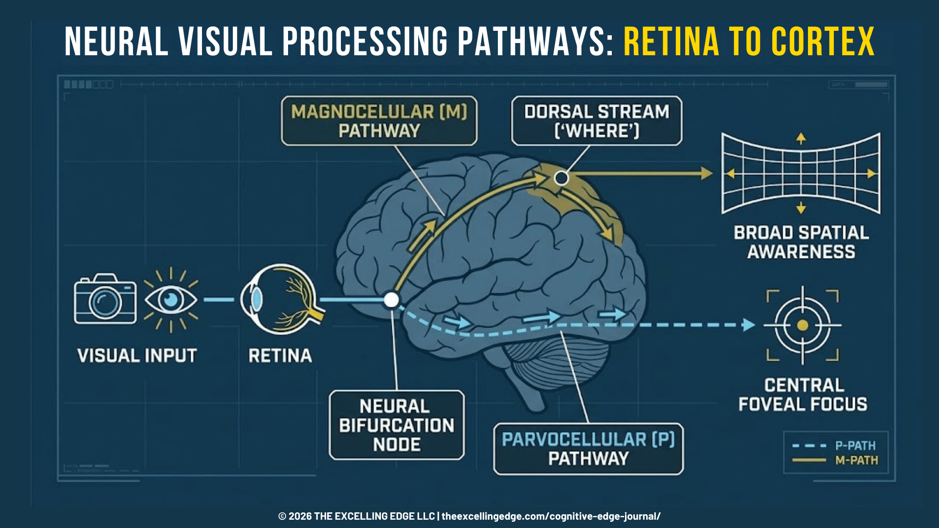
1. Parvocellular (P) Pathway — Ventral Stream (“What”)
- Anatomical Origin: Begins with P-type retinal ganglion cells and projects to the parvocellular layers of the Lateral Geniculate Nucleus (LGN), terminating in the primary visual cortex (V1) before feeding the ventral visual stream (inferior temporal cortex).
- Functional Role: Responsible for high spatial frequency, fine detail, color perception, and central foveal fixation.
- Temporal Characteristics: Slow processing speed (>100 ms processing delay); visual input must be stationary or moving slowly for accurate resolution.
2. Magnocellular (M) Pathway — Dorsal Stream (“Where / How”)
- Anatomical Origin: Begins with M-type retinal ganglion cells and projects to the magnocellular layers of the LGN, feeding directly into the dorsal visual stream (area V5/MT and posterior parietal cortex).
- Functional Role: Responsible for low spatial frequency, motion detection, spatial orientation, optic flow, and peripheral awareness.
- Temporal Characteristics: Ultra-fast transmission speed (30 – 50 ms delay); optimized for rapid spatial localization and subconscious interception.
What Drives Peripheral Vision Loss Under Stress?
When an athlete experiences intense physical exertion or acute competition stress, the Locus Coeruleus-Norepinephrine (LC-NE) system increases its tonic firing rate (Aston-Jones & Cohen, 2005). Moderate norepinephrine discharge enhances the signal-to-noise ratio in sensory processing. However, hyper-arousal and excessive norepinephrine release fundamentally alter cortical visual processing:
- Attentional Field Narrowing: High sympathetic tone triggers selective suppression of dorsal stream cortical areas. The central nervous system prioritizes central visual targets (foveal focus) to lock onto perceived primary threats, effectively dampening input from the peripheral visual fields (Easterbrook, 1959; Janelle, 2002).
- Superior Colliculus & Retinotectal Suppression: The superior colliculus, which coordinates rapid saccadic eye movements and peripheral target engagement, experiences altered top-down control from the prefrontal cortex (PFC). Subcortical survival networks take over, locking gaze onto central stimuli and increasing latency for peripheral visual acquisition.
- Cortical Receptive Field Shrinkage: Electrophysiological data demonstrates that under extreme autonomic arousal, visual cortical neurons servicing the far periphery (>30° visual angle) experience reduced receptive field sensitivity. The effective visual field shrinks from a normal 180° horizontal span to a narrow visual cone of 15° to 30°.

Lab Note: The Neuro-Visual Bottleneck
In-game “tunnel vision” is not an ocular muscle failure; it is a central nervous system compensation. Under severe metabolic strain (HR > 85% HR max), hyper-activity in the Locus Coeruleus suppresses magnocellular (dorsal stream) processing. This reduces functional peripheral awareness by up to 45% while preserving central foveal focus.
Field Diagnostics: Quantifying Visual Degradation Above 85% HR max
At our Neuro Performance Center in Surprise, AZ, we assess visual field degradation under physical stress across elite athletes from the NFL, NCAA, and tactical sectors.
During baseline assessments using digital visual testing systems (such as the Senaptec Sensory Station), elite athletes demonstrate high visual processing speed and peripheral target acquisition at resting heart rates (HR < 60 bpm). However, when subjected to progressive metabolic stress protocols (utilizing air bikes or high-velocity agility circuits to push heart rates above 85% HR max), their peripheral spatial processing metrics degrade significantly.
Diagnostic Observations (Surprise, AZ Center)
- Peripheral Latency Degradation: Reaction speed to visual stimuli beyond 30° of visual eccentricity slows down by an average of 38% under high metabolic load.
- Saccadic Overshoot (Hypermetria): Uncontrolled sympathetic arousal degrades ocular motor precision. Athletes exhibit frequent saccadic overshooting when attempting to re-fixate from central targets to peripheral cues.
- Peripheral Omission Errors: Under max-effort fatigue, athletes completely miss up to 40% of transient peripheral targets presented in their outer visual fields (>45°).
[insert graphic: NEURO-VISUAL DECAY UNDER METABOLIC STRESS]

To eliminate this bottleneck, performance directors cannot rely on static vision exercises or off-field ocular tracking. Training protocols must force the visual cortex to maintain magnocellular stream integration while operating under elevated catecholamine levels and physical fatigue.
Operational Interventions
We utilize dynamic visual occlusion and gaze anchoring protocols under metabolic load to train these systems (Appelbaum & Erickson, 2018).
- Liquid Crystal Strobe Occlusion: Strobe eyewear alternates between transparent and opaque states at adjustable frequencies (e.g., 4 Hz down to 1 Hz). By reducing visual information to brief snapshots, strobe occlusion forces the brain to speed up visual processing, rely on internal predictive models, and maximize magnocellular motion detection.
- High Activation-Visual Coupling: Strobe eyewear training is applied while athletes perform high-intensity metabolic efforts (80%-90% HR max) and execute sport-specific spatial reads.
- Visual Anchor Fixation with Peripheral Extraction: Athletes maintain central gaze lock on a primary target (e.g., keying on a central defender) while simultaneously identifying, processing, and calling out peripheral targets flashed across their outer visual fields.
The Dynamic Occlusion Framework: Expanding Spatial Awareness Under Pressure
Preserving spatial processing under stress is essential for elite athletes, tactical personnel, and high-performing corporate executives. High-stakes decision-making in executive boardrooms triggers similar sympathetic arousal spikes, restricting cognitive bandwidth and narrowing environmental awareness.
The following three-phase protocol builds neural resilience against stress-induced visual field collapse.
| Phase 1 | Phase 2 | Phase 3 |
|---|---|---|
| Gaze Anchoring | Strobe + Metabolic Load | Dual-Task Agility |
| Static Expansion | 80-85% HR max, Intermittant Occlusion | Complex Spatial Execution |
Phase 1: Static Gaze Anchoring & Visual Field Expansion
Target Population: Athletes, Executives, Tactical Operators.
Objective: Establish top-down cortical control over peripheral visual field allocation without metabolic strain.
Execution:
- Sit or stand comfortably, fixating central gaze on a stationary target 2 meters away.
- Maintain strict visual fixation on the central point. Without moving eyes or head, consciously expand visual awareness outward to detect objects, movement, and boundaries at the far left, right, top, and bottom edges of the visual field.
- Hold this broad spatial awareness state while performing controlled, slow diaphragmatic breathing (4 second inhale, 6 second exhale) to keep sympathetic arousal low.
- Volume: 30 sets x 60 seconds daily prior to performance or decision-heavy tasks.
Phase 2: Dynamic Visual Occlusion Under Metabolic Load
Target Population: High-Performance Athletes & Tactical Units.
Objective: Force dorsal visual stream processing during high autonomic arousal.
Execution:
- Equip liquid crystal strobe eyewear set to a moderate pulse rate (6 Hz).
- Initiate high-intensity cardiovascular work (assault bike, treadmill, or shuttle runs) until heart rate exceeds 80-85% HR max.
- While maintaining this target heart rate, execute reactive ball catches, target identification, or spatial decision drills using peripheral visual cues.
- Volume: 5 sets x 45 seconds work intervals 15 seconds rest, 2-3 times per week.
Phase 3: Dual-Task Cognitive-Motor Peripheral Loading
Target Population: Return-to-Play Athletes & Executive Longevity Clients.
Objective: Integrate peripheral spatial awareness with dynamic motor execution and prefrontal cognitive processing.
Execution:
- Perform dynamic movement drills (e.g., reactive shuffling or multi-directional footwork).
- Maintain central visual tracking of an oncoming trainer or visual cue.
- Verbally identify light indicators or numbered targets flashed randomly in the peripheral visual field (45° to 60° angles) without breaking physical movement rhythm or dropping central visual focus.
- Volume: 6 sets x 30 seconds with 45 seconds full recovery.
Key Takeaway: The Plasticity of Spatial Processing
Stress-induced tunnel vision is not a fixed physiological limit – it is a trainable visual processing capacity. By systematically pairing high heart rates with dynamic visual occlusion, the brain increases neural gain across the dorsal visual pathway, expanding functional spatial awareness during peak operational stress.
Optimize Your Team’s Visual Execution Under Pressure
Visual processing errors during late-game scenarios are not random mental lapses – they are trainable neuro-physiological bottlenecks. At the Neuro Performance Center in Surprise, AZ, we apply sensory diagnostic testing, strobe occlusion technology, and metabolic integration protocols to quantify and expand visual spatial awareness under peak operational stress.
Schedule an In-Center Assessment at our Neuro Performance Center to analyze your athletes’ visual processing under metabolic load and implement customized protocols to improve in-game performance.
References
- Appelbaum, L. G., & Erickson, G. (2018). Sports vision training: A review of the state-of-the-art in digital training techniques. International Review of Sport and Exercise Psychology.
- Aston-Jones, G., & Cohen, J. D. (2005). An integrative theory of locus coeruleus-norepinephrine function: Adaptive gain and optimal performance. Annual Review of Neuroscience.
- Janelle C. M. (2002). Anxiety, arousal and visual attention: a mechanistic account of performance variability. Journal of sports sciences.
- Easterbrook, J. A. (1959). The effect of emotion on cue utilization and the organization of behavior. Psychological Review, 66(3), 183–201.
- Hülsdünker, T., Ostermann, M., & Mierau, A. (2019). The speed of neural visual motion perception and processing determines the visuomotor reaction time of young elite table tennis athletes. Frontiers in behavioral neuroscience.