As a top performing executive, you operate in an environment where peak cognitive performance—your information processing, working memory, and mental agility—is your greatest asset. However, a significant blind spot exists in current executive health optimization. While you likely track your cardiovascular, metabolic, and physical metrics with precision, cognitive stamina is often relegated to a static baseline, an assumption that fails to account for the reality of neurological aging. Treating your brain as a fixed constant rather than a dynamic, trainable system is a strategy that leaves your most important competitive advantage at risk.

When cognitive fatigue inevitably degrades prefrontal processing speed, standard practice relies on central nervous system stimulants or pharmaceutical sleep aids. These interventions mask neurological taxing and exhaustion without addressing the underlying structural degradation of prefrontal cortical networks.

The prefrontal cortex (PFC) exhibits earlier and steeper age-related structural decline than motor or primary sensory cortices. Reductions in gray matter volume, synaptic density, and microvascular perfusion directly impair the neural networks responsible for top-down executive control (Raz et al., 2005).
Treating cognitive fatigue as a chemical deficiency rather than a neuroplastic conditioning gap results in accelerated burnout and compromised long-term cognitive reserve.
Preserving decision-making speed and establishing resistance to cognitive decline requires combining progressive cognitive loading with real-time autonomic nervous system regulation. Physical conditioning alone cannot shield prefrontal processing networks from degradation; targeted neurocognitive training is necessary to maintain neural plasticity across an aging executive brain.
The Science of Aging and Executive Performance
Let’s look at the neuroscience that drives executive functioning in your brain. Executive function depends on the structural integrity of the dorsolateral prefrontal cortex (dlPFC) and the anterior cingulate cortex (ACC). These structures execute working memory management, impulse inhibition, and strategic attentional allocation. Beginning in the fourth decade of life, the PFC undergoes structural shifts: dendritic spines retract, synaptic connections thin, and localized microvascular perfusion decreases (Arnsten, 2009).
Simultaneously, the locus coeruleus-norepinephrine (LC-NE) system and mesocortical dopaminergic projections experience age-related reductions in receptor sensitivity and neurotransmitter synthesis. Optimal catecholamine levels (dopamine and norepinephrine) must bind to high-affinity alpha-2A adrenoceptors and D1 receptors in the dlPFC to sustain persistent neural firing during complex cognitive operations (Arnsten, 2009).
When stress or aging disrupts catecholamine equilibrium, prefrontal microcircuits experience a reduced signal-to-noise ratio. This degradation manifests clinically as increased distractibility, information processing delays, and reduced mental flexibility under stress.

Sensory-Motor Integration and Visual-Vestibular Processing
Cognitive speed relies directly on oculomotor control and visual integration. Over 50% of the human cerebral cortex engages in visual processing, gaze stabilization, and spatial orientation. The visual-vestibular-autonomic loop operates as the primary sensory gate for executive execution:
$$\text{Retinal Input} \longrightarrow \text{Superior Colliculus} \longrightarrow \text{Frontal Eye Fields (FEF)} \longrightarrow \text{dlPFC}$$
Poor sensory input corrupts strategic decision making and performance. Age-related delays in saccadic accuracy, visual pursuit alignment, and contrast sensitivity degrade the inputs fed into the prefrontal cortex. When visual processing efficiency slows, the PFC must redirect valuable attentional bandwidth to basic spatial stabilization, reducing the metabolic energy available for high-level reasoning and risk assessment (Park & Reuter-Lorenz, 2009).
Cognitive Reserve and the Scaffolding Theory of Aging (STAC-r)
Building long-term neurological resilience requires expanding cognitive reserve—the brain’s capacity to optimize performance through alternate neural pathways when primary networks face structural strain. Under the Scaffolding Theory of Aging and Cognition (STAC-r), the brain adapts to biological changes by recruiting compensatory neural scaffolding (Park & Reuter-Lorenz, 2009).
Lab Note: Prefrontal Resource Allocation
High-performance execution requires minimizing the metabolic cost of spatial orientation. When visual-vestibular processing slows, the prefrontal cortex expends excess metabolic resources on basic environmental tracking, compromising the bandwidth needed for executive decisions under operational stress.
Targeted, non-repetitive cognitive-motor challenges stimulate this scaffolding. Subjecting the nervous system to visual-cognitive tasks while managing physical or autonomic strain triggers Activity-Dependent Synaptogenesis and systemic expression of Brain-Derived Neurotrophic Factor (BDNF) (Erickson et al., 2011). This biological cascade strengthens prefrontal networks, preserving processing speed and decision accuracy throughout the lifespan.
This doesn’t just improve short-term performance, but strengthens executive networks that support long-term cognitive capability and healthspan.
Real-World Evidence: Quantifying Executive Performance Gains
At the Neuro Performance Center in Surprise, Arizona, we evaluate tactical operators, elite athletes, and executive leaders using closed-loop diagnostic protocols. Rather than separating physical capacity from mental output, our diagnostic battery measures visual-motor integration, executive processing speed, dynamic oculomotor control, and autonomic balance under high cognitive load.
Longitudinal Diagnostic Case Data (2-Year Tracking)
To quantify the impact of systematic neurocognitive loading combined with autonomic regulation, we tracked an executive client at our Surprise, AZ center over a two-year period (December 2023 through February 2025). Diagnostics were collected on the Senaptec Sensory Station to establish percentile rankings against a normative database of elite performers.
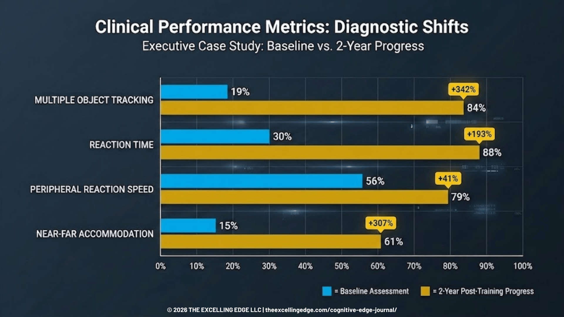
Diagnostic Shifts & Clinical Analysis:
- Multiple Object Tracking (MOT) & Dynamic Attention: In December 2023, the client scored in the 19th percentile for MOT, indicating compromised spatial tracking and attentional resources under load. Following dual-task training incorporating stroboscopic occlusion and dynamic target identification, performance reached the 84th percentile by February 2025.
- Reaction Time & Neural Efficiency: Initial assessments measured total reaction time in the 30th percentile. By February 2025, total reaction speed reached the 88th percentile (346 ms overall latency; 366 ms dominant, 325 ms non-dominant), reflecting increased transmission velocity along corticospinal pathways.
- Peripheral Processing & Oculomotor Control: Peripheral reaction speed advanced from the 56th to the 79th percentile (751 ms response latency), while Near-Far Quickness moved from the 15th to the 61st percentile. Contrast sensitivity demonstrated a parallel shift from the 5th to the 34th percentile.
These longitudinal metrics confirm that prefrontal processing networks remain highly plastic in mature adults. Systematically overloading the visual-spatial processing loop while regulating autonomic stress restores processing latency and expands functional cognitive reserve.
Lab Note: Autonomic Balance and Visual Capture
Maintaining high heart rate variability (HRV) under cognitive strain stabilizes prefrontal regulation. When sympathetic arousal overwhelms parasympathetic braking, visual search patterns become chaotic, increasing processing delays and errors in decision-making.
Operationalizing Neuro-Longevity: Your Daily Protocol
Translating these principles into daily routines requires protocols that combine autonomic control, oculomotor conditioning, and dual-task cognitive loading. This structured protocol strengthens prefrontal processing stamina and reinforces cognitive reserve.
[image 4 – protocol]
Phase 1: Autonomic Down-Regulation (Resonant Breathing) – Readiness
- Objective: Establish vagal tone and balance catecholamine release prior to high cognitive loads.
- Execution: Perform 5 minutes of resonant frequency breathing at 6 breaths per minute (4-second inhale, 6-second exhale) using real-time HRV biofeedback.
- Mechanism: Resonant breathing amplifies Respiratory Sinus Arrhythmia (RSA), increasing vagal afferent signals to the nucleus tractus solitarius and stabilizing prefrontal circuits against stress-induced performance drops (Arnsten, 2009).
Phase 2: Dynamic Visual-Oculomotor Saccades – Priming
- Objective: Drive regional blood flow to the Frontal Eye Fields (FEF) and associated prefrontal networks.
- Execution: Position two visual targets 6 feet apart on a wall. Keeping the torso stationary, perform rapid, precise saccadic eye movements between targets for 60 seconds. Transition immediately to 60 seconds of smooth pursuit tracking (moving a target in a figure-eight pattern at arm’s length while maintaining continuous central visual focus).
- Mechanism: Activates cranial nerves III, IV, and VI while stimulating frontoparietal attentional networks, raising baseline cortical readiness without elevating systemic stress hormones.
Phase 3: Dual-Task Cognitive Overload Protocol – Training
- Objective: Induce cognitive-motor interference to stimulate synaptogenesis and expand cognitive reserve.
- Execution: Combine a physical balance or movement challenge (e.g., single-leg stance on an unstable surface or multi-directional footwork) with working memory tasks (e.g., auditory 2-back tests or serial subtractions by 7s). Progress over time to introduce stroboscopic eyewear or dynamic target changes to increase visual processing demand.
- Frequency: 10 to 15 minutes per session, 3 to 4 days per week.

Cognitive resilience demands proactive conditioning. Don’t wait for noticeable slippage or symptoms to show up. To evaluate your baseline processing speed, visual-motor performance, and autonomic stress resilience, schedule an assessment with our expert human performance team.
Schedule Your In-Center Assessment at our Surprise, AZ Neuro Performance Center
References
- Arnsten, A. F. (2009). Stress signaling pathways that impair prefrontal cortex structure and function. Nature Reviews Neuroscience.
- Erickson, K. I., Voss, M. W., Prakash, R. S., et al. (2011). Exercise training increases size of hippocampus and improves memory. Proceedings of the National Academy of Sciences.
- Park, D. C., & Reuter-Lorenz, P. (2009). The adaptive brain: Aging and neurocognitive scaffolding. Annual Review of Psychology.
- Raz, N., Lindenberger, U., Rodrigue, K. M., Kennedy, et al. (2005). Regional brain changes in aging healthy adults: General trends, individual differences and modifiers. Cerebral Cortex.
- Stern, Y. (2009). Cognitive reserve. Neuropsychologia.